
Persistent Immune Dysregulation in Long COVID? A new pre-print

The preprint “Persistent Immune Dysregulation during Long COVID is Manifested in Antibodies Targeting Envelope and Nucleocapsid Proteins” by Rehman and colleagues (2025) proposes that individuals with Post-Acute Sequelae of SARS-CoV-2 infection (PASC or Long COVID) exhibit long-lasting alterations in humoral and cellular immunity characterised by elevated IgG against SARS-CoV-2 Envelope (E) and Nucleocapsid (N) proteins, skewed immune cell profiles, and an inflammatory cytokine milieu persisting up to six months post-infection. While these findings reflect a real need to understand Long COVID immunopathology, several methodological and interpretative limitations constrain the strength of the conclusions at this stage. Importantly, this is a preprint that has not yet undergone peer review.
Cohort size, heterogeneity, and clinical definition of Long COVID
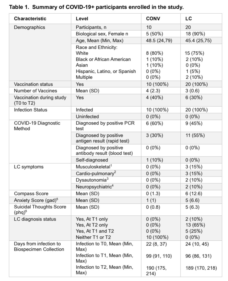
A recurring limitation in Long COVID immunology studies is small (10 vs 20) and heterogeneous cohorts with variable symptom definitions, a problem that appears here as well. PASC encompasses a broad range of clinical presentations, and grouping diverse phenotypes into a single category may mask many important mechanistic differences or create spurious associations driven by outliers. Without clear stratification by symptom clusters (e.g., fatigue versus cardiopulmonary versus neurological manifestations) or severity scales, it is difficult to generalise immunological patterns across the full spectrum of Long COVID.
Similarly, relatively modest sample sizes reduce statistical power and increase the chance that observed differences (e.g., elevated E/N IgG titters) could be artefacts of sampling variation rather than robust biological signals. This weakens a study and reduces the conclusions that can be made.
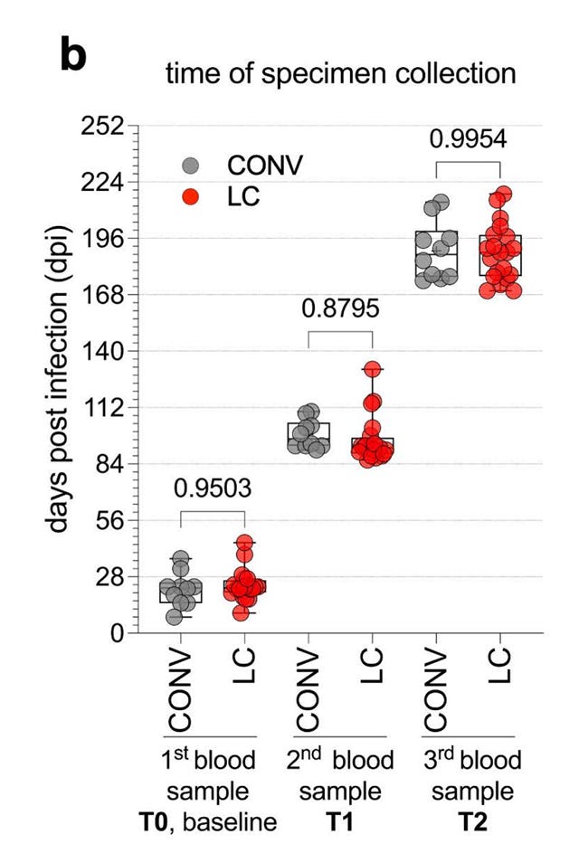
The retrospective study used the RECOVER cohort with confirmed SARS-CoV-2 infections between December 2021 and October 2022.
Peripheral blood versus tissue-specific immunity
The study focuses on peripheral blood immunoprofiling (circulating antibodies, cytokines, and immune cell subsets). While accessible, blood measurements are an imperfect proxy for tissue-level immune events, especially when mechanisms like tissue viral persistence, local inflammation, or mucosal immune responses might play key roles in pathobiology. Studies on Long COVID (and other disorders such as tissue-specific autoimmunity) have highlighted that immune phenotypes may involve tissue compartments; lungs, gut, even nervous system, which are not captured by blood assays. Consequently, peripheral immune signatures should not be overinterpreted as evidence of ongoing antigen exposure or systemic pathology without corroborating tissue data.
Interpretation of elevated antibodies
The authors argue that persistent anti-Envelope and anti-Nucleocapsid IgG responses suggest ongoing antigenic stimulation. However, several alternative explanations exist:
Antibody kinetics differ by antigen: Immune memory to certain viral proteins can naturally persist long after clearance of infectious virus without implying ongoing antigen presence. For instance, high levels of Nucleocapsid-specific antibodies have been reported to persist shorter than Spike-specific responses in some cohorts, even when levels remain detectable many months later, this can be explained by memory plasma cell and memory B cell dynamics rather than persistent antigen.
Memory B cells and long-lived bone marrow plasma cells can maintain antigen-specific IgG antibody levels for many months or longer in the absence of antigen, because plasma cells continually secrete antibody. This is well established for viral infections generally, and demonstrated in SARS-CoV-2.
Longitudinal cohorts show that SARS-CoV-2 memory B cell responses persist and evolve for many months, even when viral replication has ceased, indicating that persistent antibody titters are consistent with normal adaptive immune memory.
Many past studies emphasize that detectable IgG months after infection is typical for many viral infections, including SARS-CoV-2, and reflects immunological memory, not necessarily ongoing infection or antigen persistence.
Affinity maturation and immune memory variability: Differences in antibody subclass distribution or chronic low-level stimulation could reflect memory imprinting rather than active dysregulation.
Autoimmunity versus persistent antigen: Elevated autoantibodies or inflammatory cytokines could arise from post-infectious immune perturbations unrelated to persistent viral antigen.
Thus, equating sustained antibody titers with viral persistence is suggestive but far from definitive. Longitudinal research with tissue sampling, direct antigen detection, and functional assays is needed to disentangle these possibilities.
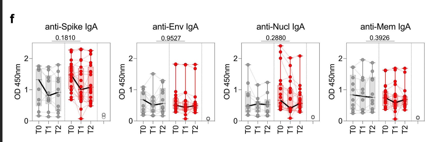
They do not find differences in IgA antibody levels in blood.
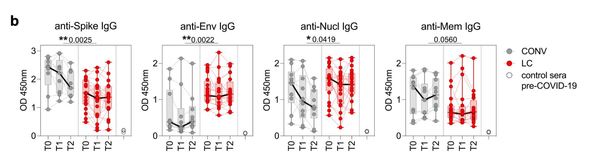
But, find differences in IgG levels. This is in contrast to many other studies, showing an increase in anti-S or anti-S1 or anti-RBD IgG levels in blood from Long COVID patients.
https://www.nature.com/articles/s41586-023-06651-y
https://academic.oup.com/ofid/article/11/4/ofae137/7638460
https://www.frontiersin.org/journals/immunology/articles/10.3389/fimmu.2025.1670324/full
https://academic.oup.com/ofid/article/11/4/ofae137/7638460
Systematic reviews and larger cohorts show that the relationship between anti-Spike antibodies and Long COVID is complex and time-dependent. Some associations indicate that lower acute anti-Spike responses may predispose to Long COVID, or that higher humoral responses correlate with particular symptom clusters, rather than a uniform elevation across all Long COVID patient.
Overall: While several studies do show higher anti-Spike IgG in Long COVID patients compared to non-LC infected controls, not all cohorts find this difference and some even report lower or no difference, depending on study design, patient demographics, time post-infection, and vaccination history. This makes it so important to have large cohorts and to stratify patient cohorts very well.
Lack of functional correlates of immune activation
While the study reports differences in cytokine levels and immune cell populations (e.g., T follicular helper cells, MAIT cells), it stops short of demonstrating functional consequences of these changes. Elevated cytokines or cell subset frequencies do not necessarily imply pathogenic activity, many are part of normal immune dynamics following infection. In the context of Long COVID, distinguishing adaptive compensatory responses from maladaptive dysregulation requires functional assays (e.g., antigen specificity of T cells, cytokine bioactivity, immune activation/inhibition markers) that go beyond descriptive immunophenotyping.
What the authors show are proportions of T cell subsets. Proportions are difficult to interpret, but seem to indicate a heightened T cell activity state (increase proportion of memory subsets). Dendritic cells (mDC, pDC), other myeloid subsets, NK and gdT cells and ILCs seem similar between controls and patients. Only an increase in mucosal associated innate T cells (MAIT) was observed.
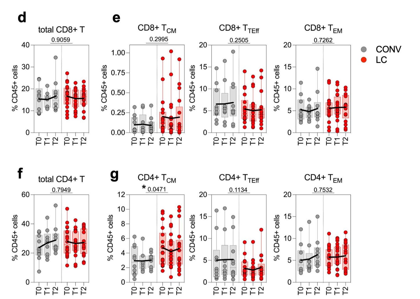
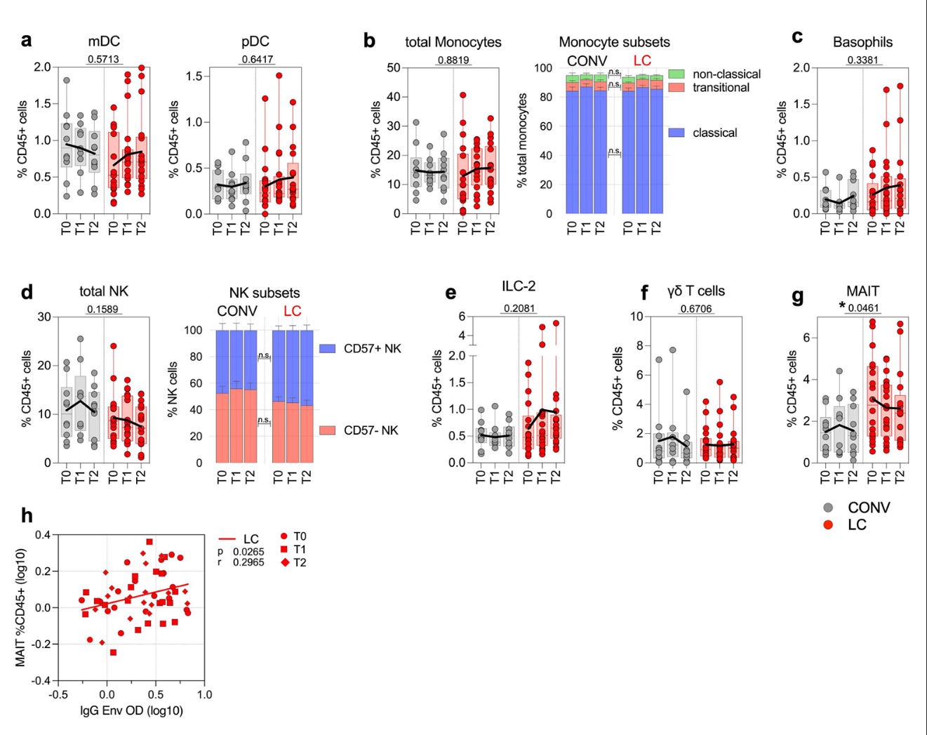
Some samples seem to show increase self-reactivity. But again, this was not seen in larger cohorts.
Time window and longitudinal stability
The six-month observation period, while helpful, may not capture longer-term trajectories of immune responses. Some studies show that immune perturbations in Long COVID evolve over 12–24 months, with possible attenuation or normalisation over time. Others link immune alterations to reinfections, vaccination, or other environmental exposures, confounding attribution to the original SARS-CoV-2 infection. Without extended follow-up and repeated sampling, cross-sectional snapshots risk overestimating the persistence and relevance of specific immune signatures.
Broader context: long COVID immunology is complex and multifactorial
The literature on post-acute SARS-CoV-2 immunology illustrates a heterogeneous landscape:
Systemic inflammation and T cell activation have been observed in multiple cohorts, yet the patterns vary by study and patient subgroup.
Some research indicates altered immune activation may improve over time, challenging models of permanent or static immune activation in all individuals.
Evidence of tissue antigen persistence remains mixed and difficult to distinguish from immunological memory without direct viral detection. Antigen may persist for longer, and some studies show RNA and peptides, but this can have alternative causes, such as decreased clearing.
Taken together, these studies suggest that persistent immune activation signatures in Long COVID are real but nuanced, shaped by factors such as viral dose, host genetics, comorbidities, and vaccination history. No single immune marker has yet emerged as a mechanistic driver or diagnostic tool.
In summary:
The Rehman et al., preprint contributes longitudinal immunoprofiling data and emphasis the known hypotheses about altered humoral responses in Long COVID patients. However, its conclusions about “persistent immune dysregulation” reflecting ongoing antigen exposure or direct pathogenic processes are premature and overinterpreted without more rigorous cohort characterisation, functional validation, and peer review. Moving from descriptive associations to causal mechanisms (what is a consequence of Long COVID and what is causing Long COVID/symptoms) will require careful integration with broader longitudinal and tissue-based studies.
As the PASC/PIAS field matures, claims about immune persistence and therapeutic targets will require much more work. With the COVID-19 pandemic there arose a unique opportunity to look at PIAS from a determined pathogen and time. ME/CFS research has encountered huge difficulties with unknown starting time and pathogen. However, now we are post-pandemic and testing for SARS-CoV-2 infection is limited, this opportunity is fading quickly, but for historical records such as RECOVER as used here.