
Inflammatory Milieu and Specific T-Cell Response Observed Three Months and One Year After SARS-CoV-2 Infection in Long COVID Subjects
Another MDPI journal publication

The authors have a very modest record in the field of immunology, some works on HCV, HIV, measles. Observational and descriptive studies are the majority of published works.
Straight to the methods section, what did they do, how did they do it?
“This is a prospective, case–control study enrolling 196 unvaccinated patients consecutively evaluated at the post-COVID clinic of the Italian National Institute for Infectious Diseases “Lazzaro Spallanzani.” Participants were enrolled between May and June 2020 and were referred either from the hospital or from community healthcare services.“ The total number of patients is 196, none were vaccinated and they were infected during the first wave.
“Of the 196 participants, 119 (60.7%) were male and 77 (39.3%) female. The median age was 56.5 years (interquartile range [IQR], 49.5–64.9)”. Important to keep in mind.
In addition, “Among the participants, 147 (75%) had previously been hospitalized for COVID-19 at our institute, while 18.9% had not required hospitalization.“ This also puts the results in context: people past 50 years of age, who contracted SARS-CoV-2 and ended up in hospital. This context means, these results any apply to similar cohorts: no prior immunity, over 55 years of age, mainly men, and mostly severe disease.
In addition, “Regarding comorbidities, 11.7% of patients reported a history of chronic respiratory disease, while 32.7% did not. However, detailed information on pre-existing conditions was not consistently available, as the clinical implications of long COVID were not yet fully recognized at that time.“ many have other health issues that may contribute and not all are known. Looking at the medication of the subjects: “This pattern reflects the high prevalence of cardiovascular comorbidities in the cohort, with frequent use of beta-blockers, ACE inhibitors, antiplatelet agents, and calcium channel blockers“ These will influence assessments of serum values.
“Patients were assigned to the long COVID group (Ongoing LC, Ong LC) if they presented with at least one symptom at the first visit (T3M), and to the asymptomatic (Never LC, Nev LC) group if they did not“ Which were those symptoms?
“fatigue, concentration or memory deficits, reduced exercise tolerance, dyspnea, arthralgia, myalgia, dysautonomia, neuropsychiatric symptoms (e.g., insomnia, low mood, nervousness), and loss of smell or taste.“ A issue here is that many are frequently found, independent of SARS-CoV-2 infection and only one is sufficient to be classified as having LongCovid (“(68.2%) reported a single symptom“). The risk is high that the LongCovid signal, if present, is missed among the background noise signals.
Important: “The two study cohorts were followed up for 12 months (T12M). After one year post infection, none of the enrolled patients showed symptomatology (Resolved LC, Res LC).” So all symptoms were gone between 3 months and 12 months.”
What did the authors study? “Blood samples (plasma and cells) were collected at three months (T3M) and twelve months (T12M) after SARS-CoV-2 infection.“
“The median time between the first reported positive SARS-CoV-2 test and the baseline ambulatory follow-up visit was 86 days (interquartile range, 78–91 days). All participants were experiencing their first episode of COVID-19.“ This means that on average, blood sample 1 was taken nearly 3 months after being infected.
Important as well, only “48 patients completed their evaluation“ at 12 months. These may not be representative of the whole cohort, nearly 4 times this size.
I will skip the tests done, these may be relevant when looking at the results.
“at T3M, no differences were observed in the levels of D-Dimer (Figure 1A–C), E-Selectin (Figure 1D–F), ICAM-1 (Figure 1G–I), and VCAM-1 (Figure 1J–L) between asymptomatic (Never LC) and symptomatic LC (Ongoing LC) subjects”
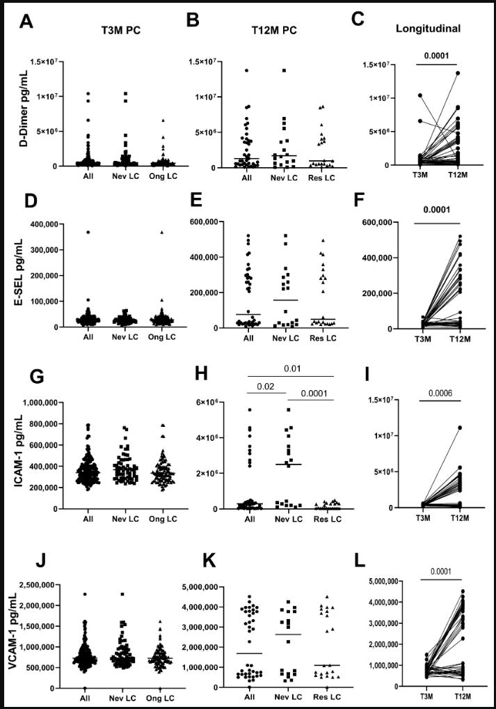
There were difference depending on severity of the disease, as you would expect: “we found that levels of ICAM-1 (p = 0.006, Figure 1G) and VCAM-1 (p = 0.02, Figure 1J) were higher in hospitalized subjects compared to their non-hospitalized counterparts, indicating greater endothelial activation “
“When comparing values from T3M to T12M within the available data, we found that levels of ICAM-1 (p < 0.0001) were higher in Never LC subjects at T12M compared to those in Resolved LC (Figure 1H). No differences were observed for the other factors between Never LC and Ongoing LC subjects.“ Have a good look at panel 1H, note in two conditions there appears a second group with higher ICAM-1 levels. Is that related at all to SARS-CoV-2 infection? There is no evidence for this.
But, “analyzing the data in a longitudinal course, we observed a significant increase in D-Dimer, E-Selectin, and VCAM-1 (p < 0.0001 for all, Figure 1C,F,L), along with a substantial rise in ICAM-1 (p < 0.0006, Figure 1I),“ When the authors compared 3 and 12 month values, all values had gone up, in most subjects.
What are normal levels?
D-dimer: <500 ng/mL = < 500.000 pg/mL
E-SEL: 1-40 ng/mL = 1-40.000 pg/mL
ICAM-1: 100-200 ng/mL = 100-200.000 pg/mL
VCAM-1: 553 ng/mL = 553.000 pg/mL
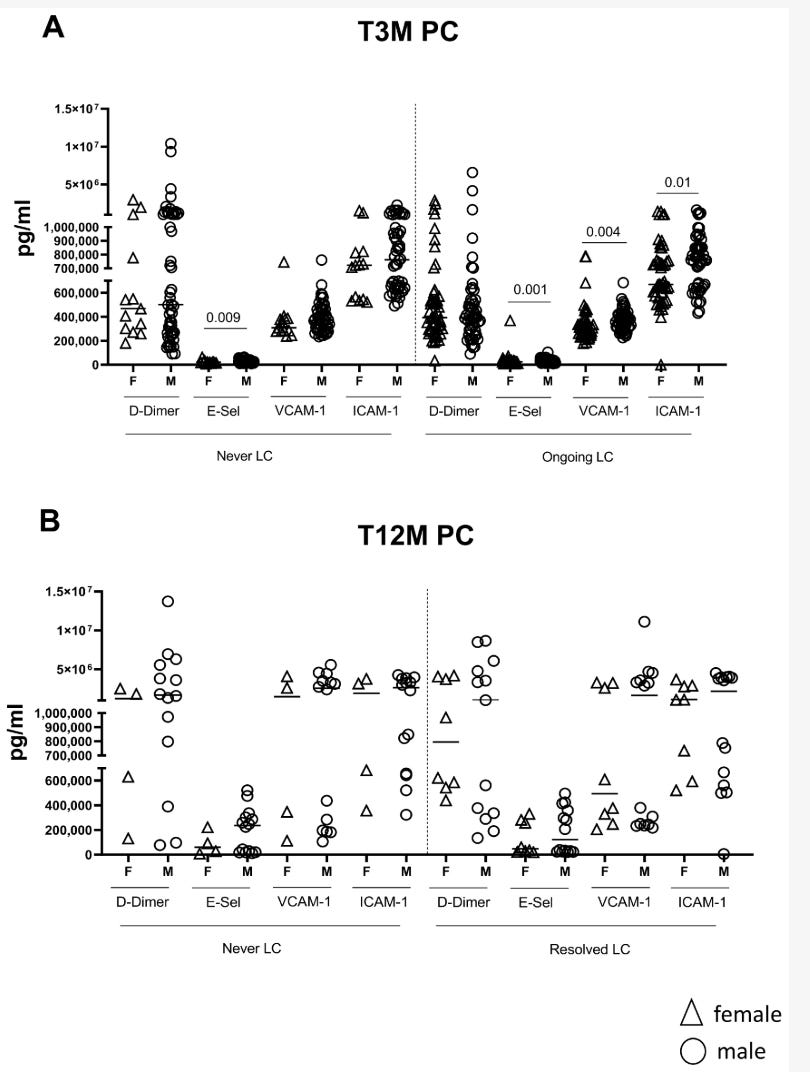
Looking at the graphs again, two things are noted, using ICAM as the example:
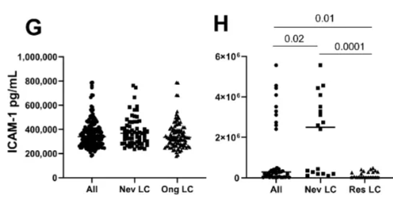
The left graph is at 3 months, the Y-axis to to 1.000.000. The right graph, this axis goes to 6.000.000. This is largely needed for those patients in a subgroup with high values. Then we look at the next panel where 3M and 12M are directly compared.
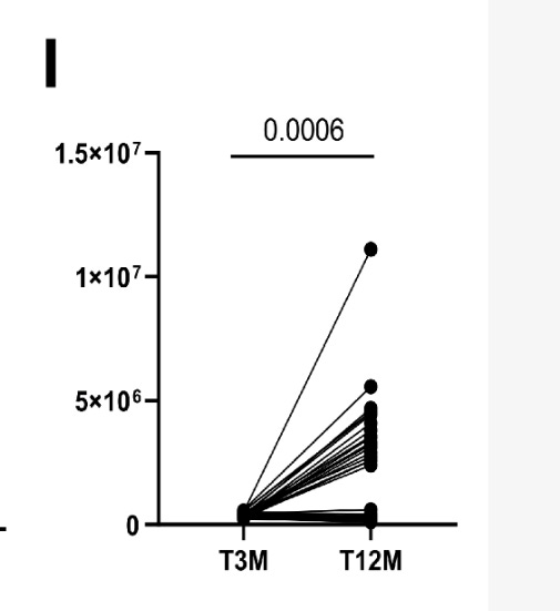
Note this axis goes to 15.000.000? Note how one sample is at at ~11.000.000? Where is that sample in panel H, none of which go over 6.000.000? Are those 25% returning at 12 moths representative? There seems a large enrichment of those with the high values, only observed in non-infected and infected at 12 months. Note also how those with high values in the non-infected controls are present as well? These are not shown, but a similar graph could be made with those. Is a conclusion about COVID-19 or damage still justified? No, it is not.
Look at D-dimers, E-SEL and VCAM, and you can see the same problem with those graphs. In addition, the second issue, are the values, some are way off of what you would expect. The difference, controls and infected, between 3 and 12 months makes me wonder if different people performed these assays on different days, or if samples were frozen from the 3month batch, etc. Something technical is concerning here as well.
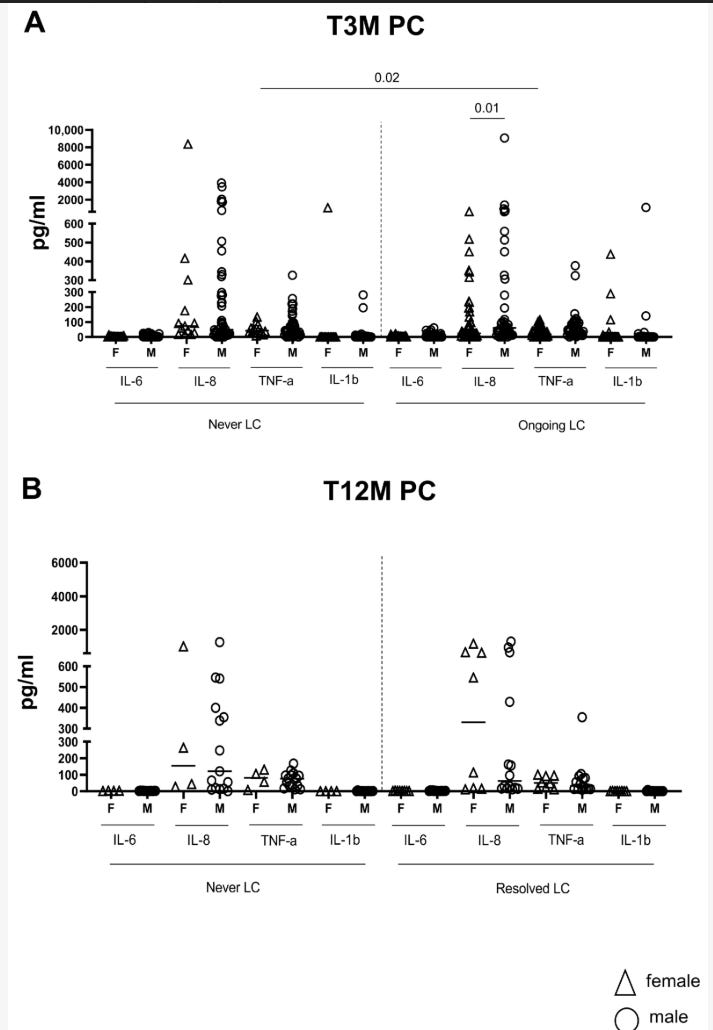
“The inflammatory profile revealed that no differences were observed for all cytokines in Never LC and Ongoing LC/Resolved LC subjects at T3M (Figure 2A,D,G,J) and T12M (Figure 2B,E,H,K).“ “the analysis of the inflammatory profile at T3M revealed higher levels of IL-6 in hospitalized patients compared to non-hospitalized (p = 0.01, Supplementary Figure S2A).“ “No group differences were observed between groups for IL-1β (p = 0.51, Supplementary Figure S2), IL-8 (p = 0.11, Supplementary Figure S2), and TNF-α (p = 0.36, Supplementary Figure S2).“
All this is expected.
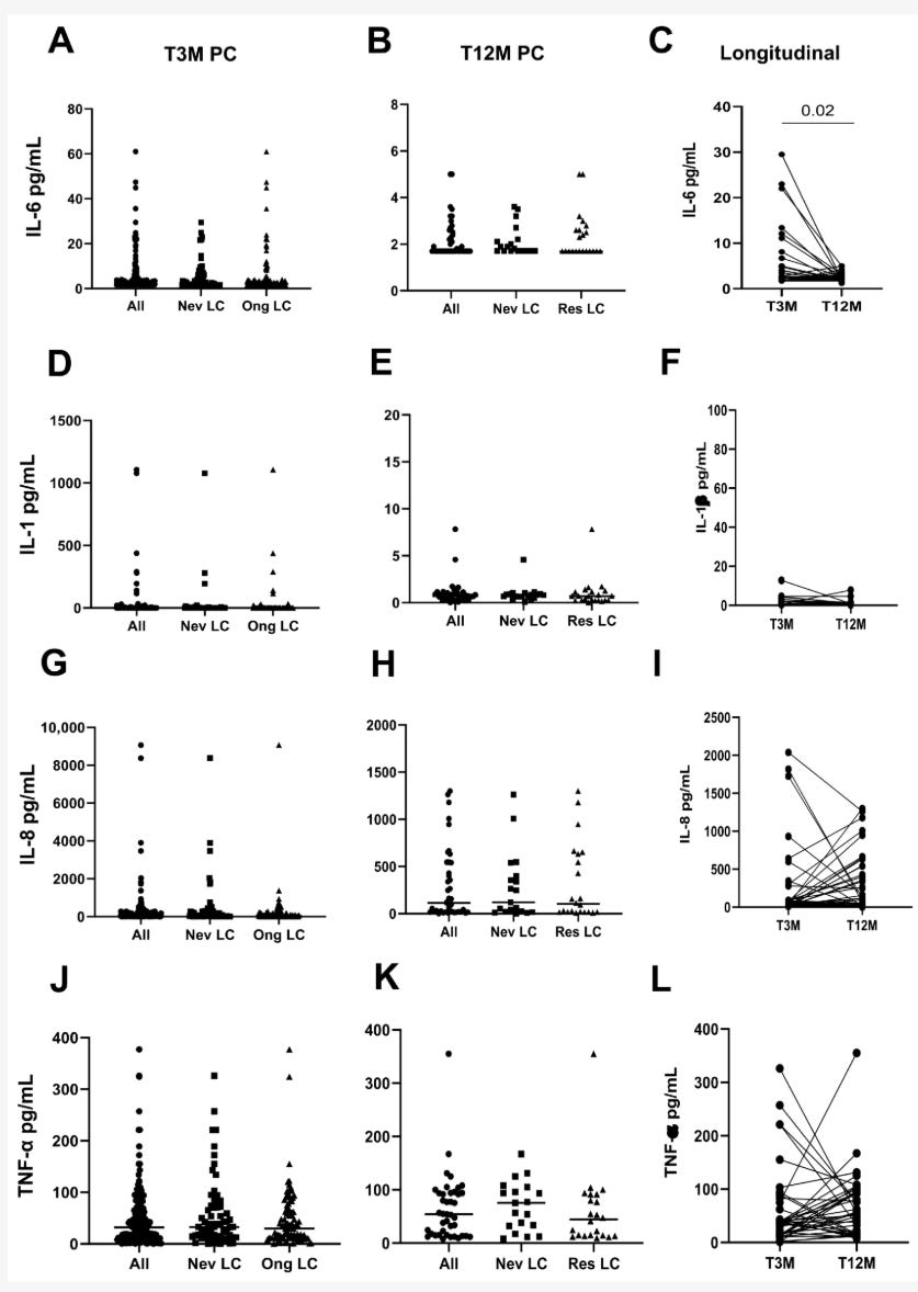
“When comparing T3M with the T12M mark, IL-6 levels experienced a significant decline over time (p = 0.02, Figure 2C), while IL-1β (p = 0.30, Figure 2F), IL-8 (p = 0.17, Figure 2I), and TNF-α (p = 0.13, Figure 2L) showed no notable changes.“ Sure, many had severe disease, IL-6 levels may still have been higher at 3 months, and these reduce back to baseline. Note again the differences on the Y-axis and the presence of some subjects with higher values within the groups, importantly: including in the non-infected.
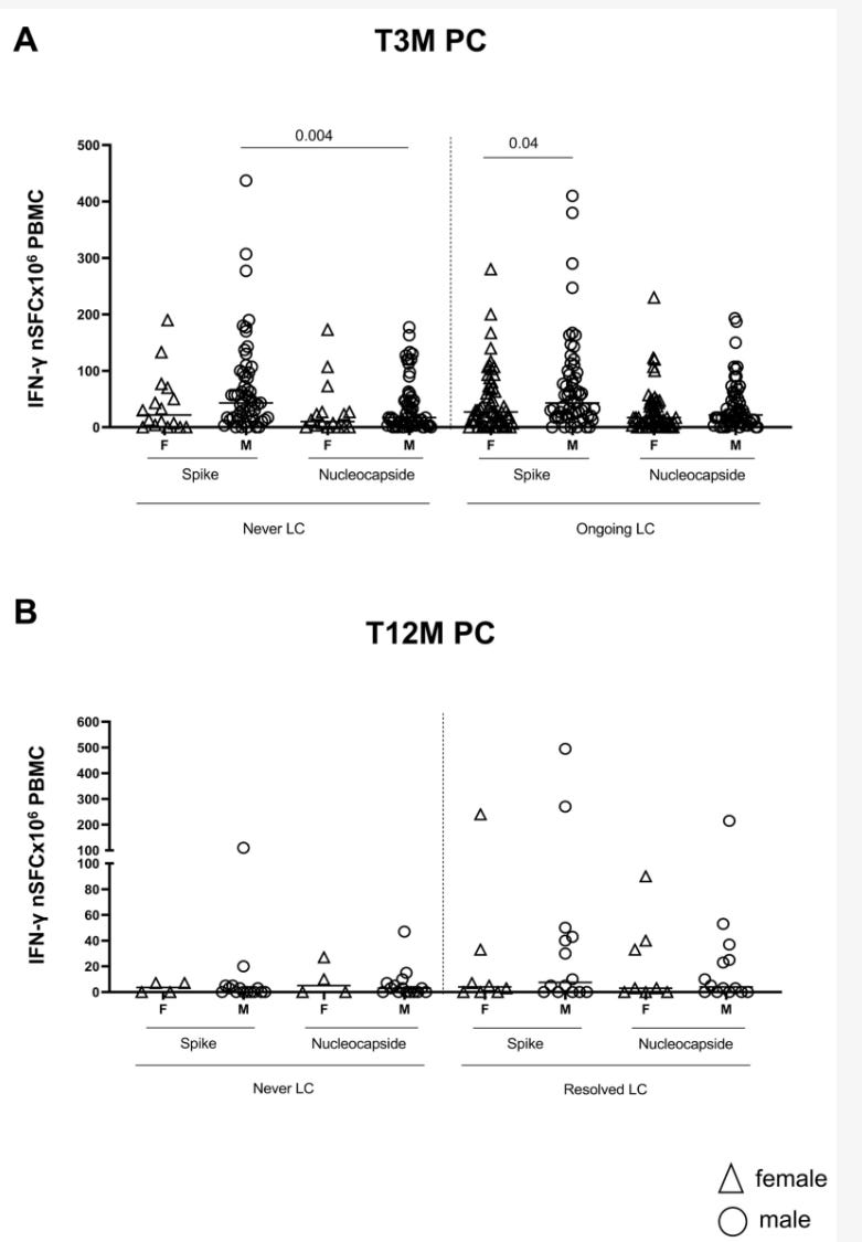
“Lastly, our data showed that there is no difference in the strength of the spike (Figure 3A,C) and nucleocapsid T (Figure 3B,D) response between Never LC and Ongoing LC at T3M and Resolved LC at T12M. At the same time, we demonstrated that the spike–SARS-CoV-2 specific T-cell response was significantly higher in hospitalized patients compared to non-hospitalized patients (p = 0.004, Supplementary Figure S3A) and also declined markedly at the 12-month mark (T12M) in the hospitalized patient’s group (p = 0.0001, Supplementary Figure S3A). “ Fine.
But, “Overall, a significant reduction in spike-specific T-cell response was observed across the cohort (T12M, p = 0.001, Figure 3C and Supplementary Figure S3C).“ should be interpreted as physiological. At 3 months there will still be effector T cells as more effector memory T cells. Many have died by 12 months (they are supposed to) and memory cells specific for SARS-CoV-2 are reduced in the blood, increased in other tissues, such as the lungs, lymph nodes, and bone marrow.
Then, which is a valid analysis, the authors compared sexes “within the LC group, male patients (n = 50) exhibited significantly higher expression of several crucial endothelial factors compared to female patients (n = 13): E-Sel (p = 0.001), VCAM-1 (p = 0.004), and ICAM-1 (p = 0.01, Figure 4A); D-Dimer expression remained comparable between LC females and males (p = 0.61, Figure 4A). However, these differences did not persist at T12M (Figure 4B).“ At 12 months there is a selection of subjects, and there are very few women remaining. Care has to be taken to draw any conclusions from this. Here it is again very visible that the returning groups is not representative of the starting group. This largely invalidates any comparison between 3 and 12 months.
“The inflammatory profile was strikingly characterized by elevated TNF-α levels in female patients with Ongoing LC (N = 49) compared to their Never LC counterparts (N = 13; p = 0.02, Figure 5A). “ Seriously?? I have no idea what the authors saw, but I do not see it. Nothing striking, not much TNF.
“Regarding adaptive immunity, the spike-specific T-cell response was similar between Ongoing LC and Never LC female patients (spike Ongoing LC vs. Never LC females = 0.52) and male patients (spike Ongoing LC vs. Never LC males = 0.88). However, a significantly stronger spike response emerged in male LC patients when compared to females (p = 0.04), underscoring the gender differences in immune response. “
So, in both, those with LC and without, the T cell response is good. No, no “immune-damage” and that sort of thing. Actually a stronger response in those reporting at least one LC symptom.
To conclude: there are issues that severely undermine any conclusions from this work.
Please don´t use it.

553 ng/mL553 ng/mL
553 ng/mL

553 ng/mL553 ng/mL
553 ng/mL